Gamma Radiograph
I find it interesting experimenting with different parts of the electromagnetic spectrum,
so I decided to try to make a gamma radiograph. In order that the image be reasonably sharp
it is necessary to use something approximating to a point source.
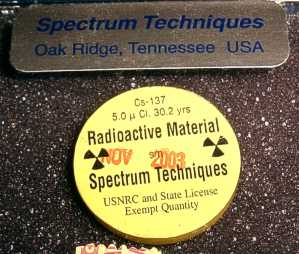 The most intense source I have which is of suitable dimensions is a 5μCi Cs137 source from
Spectrum Techniques.
It was purchased legally as it is a license exempt sealed source. The disc is 25mm in
diameter, but the actual source inside is only 5mm in diameter (determined by X-ray).
The most intense source I have which is of suitable dimensions is a 5μCi Cs137 source from
Spectrum Techniques.
It was purchased legally as it is a license exempt sealed source. The disc is 25mm in
diameter, but the actual source inside is only 5mm in diameter (determined by X-ray).
The main gamma peak of Cs137 is at 662keV. This makes it very penetrating. Unfortunately,
being only 5μCi I calculated that the exposure time would need to be at least two months.
In practice the situation is even worse because of reciprocity failure of the film for such
a long exposure time.
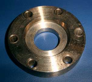 This is the item which I decided to use as the object to be radiographed. It is a stainless
steel vacuum flange with an overall diameter of 70mm. The outer annulus is 12.6mm thick and
the inner annulus is about 6mm thick.
This is the item which I decided to use as the object to be radiographed. It is a stainless
steel vacuum flange with an overall diameter of 70mm. The outer annulus is 12.6mm thick and
the inner annulus is about 6mm thick.
I chose this item because I did not expect to obtain a very clear image and this shape would be
easy to recognize and also provides some variation in thickness. It is also far too absorbing
to X-ray at 50keV.
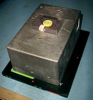 I loaded a sheet of Agfa Structurix D4 X-ray film into an Agfa Curix cassette with green
intensifier screens. On top of the cassette I placed the vacuum flange and a 100mm high
die-cast aluminium box. I then placed the Cs137 source on top of the box.
I loaded a sheet of Agfa Structurix D4 X-ray film into an Agfa Curix cassette with green
intensifier screens. On top of the cassette I placed the vacuum flange and a 100mm high
die-cast aluminium box. I then placed the Cs137 source on top of the box.
I put the whole thing in a cupboard and left it alone for two months. The exposure was
started at 17:20 hours on the 19th of December 2004 and was stopped at 19:40 hours on
the 19th of February 2005. The film was then developed using ordinary black and white
photographic paper developer. Developing time was 4 minutes and fixing time was 2
minutes.
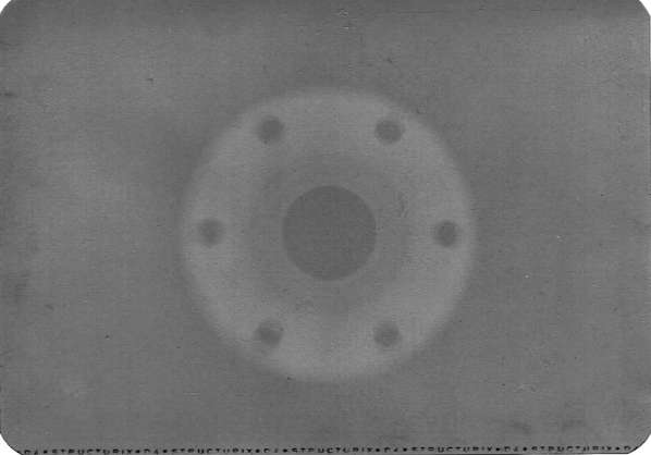
The image below is a scan of the developed film. The contrast has been boosted a great
deal. The image on the film is very faint. The middle annulus of the flange has been
penetrated enough to cause significant exposure. This part of the flange is 6 to 7mm
thick stainless steel. The 50keV X-rays used for the radiographs in the
X-Ray Gallery
would not have penetrated even 1mm of steel.